Introduction
Fusion biopsy is an advanced medical procedure that combines imaging techniques to improve the accuracy of tissue sampling for diagnostic purposes. It is primarily used in the detection of prostate cancer but is also being explored for other medical applications. By integrating magnetic resonance imaging (MRI) with real-time ultrasound, fusion biopsy enhances the precision of targeting abnormal tissues, increasing the likelihood of accurate diagnosis and effective treatment. This article explores the benefits, procedure, and applications of fusion biopsy.
Definition
Fusion biopsy is an advanced diagnostic technique that combines magnetic resonance imaging (MRI) with real-time ultrasound to improve the accuracy of tissue sampling, particularly in detecting prostate cancer. This method enhances the precision of targeting suspicious areas, reducing the risk of missed diagnoses compared to traditional ultrasound-guided biopsies.
What is Fusion Biopsy?
Fusion biopsy, also known as MRI-ultrasound fusion biopsy, is a diagnostic technique that integrates MRI scans with ultrasound imaging to guide the biopsy needle precisely to areas of concern. This hybrid approach improves the detection of suspicious lesions compared to traditional biopsy methods, which rely solely on ultrasound guidance.
Traditional ultrasound-guided biopsies can sometimes miss tumors or fail to detect aggressive cancerous cells, leading to false negatives or misdiagnosis. The fusion biopsy method overcomes these limitations by leveraging MRI’s high-resolution images to map the prostate or other targeted organs in greater detail, enabling physicians to pinpoint abnormal tissue with greater accuracy.
Benefits of Fusion Biopsy
Increased Accuracy and Early Detection:
One of the most significant advantages of fusion biopsy is its ability to accurately detect cancerous lesions at an early stage. By combining MRI with ultrasound, doctors can precisely target suspicious areas, reducing the risk of missing aggressive cancer cells. This leads to better patient outcomes through timely diagnosis and treatment.
Minimally Invasive Procedure:
Fusion biopsy is a minimally invasive procedure, meaning it requires only a small needle to extract tissue samples. Compared to surgical biopsy methods, it reduces patient discomfort, lowers the risk of complications, and shortens recovery time.
Reduced Need for Repeat Biopsies:
Because of its enhanced precision, fusion biopsy minimizes the likelihood of false negatives, reducing the need for repeat biopsies. Patients can receive a more definitive diagnosis in a single session, saving time and avoiding unnecessary procedures.
Improved Risk Stratification:
Fusion biopsy not only detects cancer but also provides a more accurate assessment of tumor aggressiveness. This allows physicians to differentiate between low-risk and high-risk cases, helping to determine the most appropriate treatment plan, whether it be active surveillance or immediate intervention.
Enhanced Patient Confidence and Peace of Mind:
A more precise and reliable biopsy process gives patients greater confidence in their diagnosis and treatment plan. The reduced uncertainty associated with traditional biopsy methods can alleviate anxiety and help patients make informed decisions about their health.
The Fusion Biopsy Procedure
The fusion biopsy procedure typically involves the following steps:
Pre-Biopsy MRI Scan:
The process begins with an MRI scan of the target organ, commonly the prostate. The MRI creates detailed images that help identify suspicious lesions or areas that require further investigation.
Image Processing and Fusion:
Specialized software is used to overlay the MRI images onto real-time ultrasound images. This fusion of imaging techniques enables doctors to visualize and navigate the area with greater accuracy during the biopsy.
Biopsy Needle Guidance:
With the help of the fused images, a biopsy needle is precisely guided to the identified lesion. The needle extracts small tissue samples from the suspicious area for further examination under a microscope.
Pathological Analysis:
The collected tissue samples are sent to a laboratory for pathological analysis. The pathologist examines the samples to determine the presence of cancerous cells, their grade, and other relevant characteristics.
Diagnosis and Treatment Planning:
Based on the biopsy results, the doctor will discuss the findings with the patient and recommend appropriate next steps. If cancer is detected, the patient may undergo further testing or begin a personalized treatment plan.
Applications of Fusion Biopsy
1. Prostate Cancer Diagnosis:
Fusion biopsy is most commonly used in the diagnosis of prostate cancer. The prostate is a small gland that can be challenging to assess using traditional biopsy methods alone. MRI-ultrasound fusion biopsy has significantly improved the detection of clinically significant prostate cancer, allowing for better patient management.
2. Detection of Other Cancers:
While fusion biopsy is primarily used for prostate cancer, researchers are exploring its application in diagnosing other cancers, such as kidney, liver, and breast cancer. The ability to integrate MRI with ultrasound for targeted tissue sampling holds promise for improving early cancer detection in various organs.
3. Evaluation of Suspicious Lesions:
Fusion biopsy is not limited to cancer diagnosis. It can also be used to evaluate suspicious lesions, cysts, or abnormalities in different organs. This helps in determining whether a lesion is benign or malignant, guiding further treatment decisions.
4. Monitoring Disease Progression:
Patients with known cancerous lesions or pre-cancerous conditions can benefit from fusion biopsy for ongoing monitoring. By periodically assessing changes in tissue characteristics, doctors can determine whether the disease is progressing, stable, or in remission.
5. Personalized Medicine and Targeted Therapy:
The detailed imaging and precise sampling provided by fusion biopsy enable a more personalized approach to treatment. By understanding the molecular and genetic characteristics of tumors, doctors can tailor treatment plans, including targeted therapies, to maximize effectiveness while minimizing side effects.
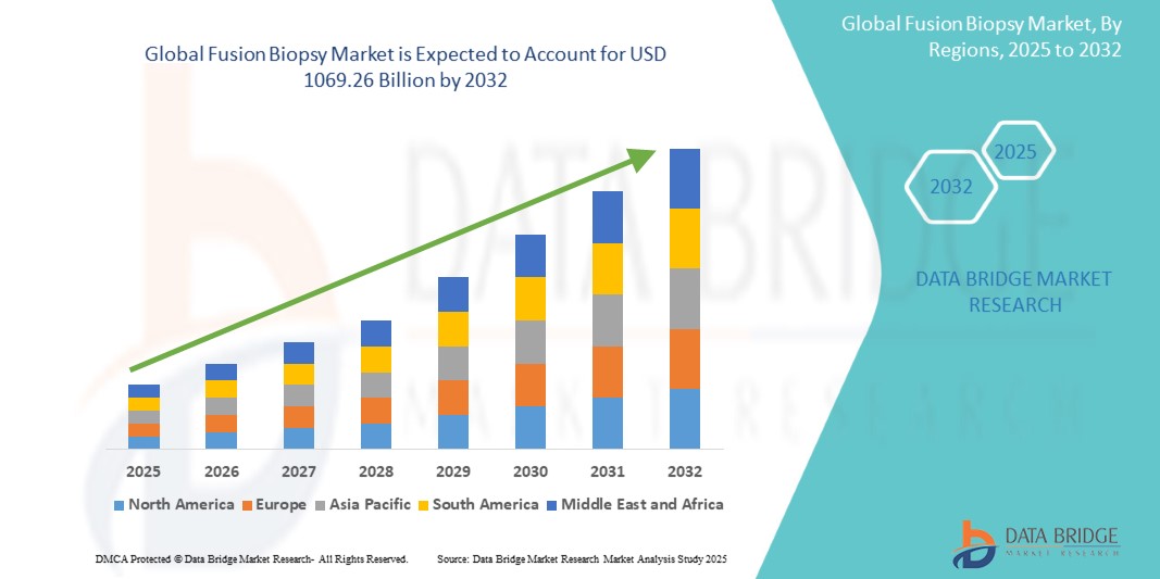
Growth Rate of Fusion Biopsy Market
According to Data Bridge Market Research, the fusion biopsy market is expected to grow at a compound annual growth rate (CAGR) of 6.40% from 2025 to 2032, from its 2024 valuation of USD 587.85 million to USD 1069.26 billion.
Read More: https://www.databridgemarketresearch.com/reports/global-fusion-biopsy-market
Conclusion
Fusion biopsy represents a significant advancement in medical diagnostics, particularly in the field of cancer detection. By integrating MRI and ultrasound imaging, it enhances the accuracy of tissue sampling, reduces the risk of false negatives, and improves overall patient outcomes. As research continues, the applications of fusion biopsy are expected to expand beyond prostate cancer to other areas of medicine, offering hope for better diagnostics and more effective treatments.


Leave a Reply